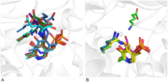
Figure 5. Comparison of fold IV and fold I transaminases.
A: Position of lysine relative to PLP in fold IV transaminases: in AT-ωTA (green), BCAT from human (1KT8, blue) or E. coli (1IYE, turquoise) and D-ATA from Bacillus sp. YM-1 (3DAA, brown). Ligands: blue: L-Ile-aldimine bound in human BCAT, turquoise: L-Glu-aldimine bound in BCAT from E. coli, brown: D-Ala-aldimine bound in D-ATA, green: L-Glu-aldimine bound in AT-ωTA, purple: docked acetophenone-aldimine in AT-ωTA. B: Position of lysine relative to PLP in fold I (S)-ω-transaminases: in PD-ωTA from Paracoccus denitrificans (4GRX, light green), PA-ωTA from Pseudomonas aeruginosa (4B98, turquoise) and several (S)-TAs identified from the Pdb by Steffen-Munsberg: from Pseudomonas putida (3A8U, pink), from Mesorhizobium loti (3GJU, yellow) and from Silicobacter pomeroyi (3HMU, brownish), in PD-ωTA the substrate, 5-aminopentanoate, is depicted in light green. The figures were prepared using the program PyMOL.

