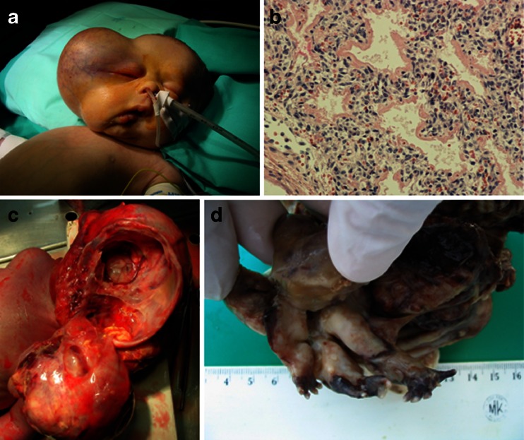
Fig. 2.
a The child prepared for the surgical removal of the tumor of the right aspect of the cranium, enormously disfiguring the child’s head. b The lung of the “host fetus” with the features of immaturity, atelectasis, and distinct hyaline membranes. Objective magnification ×20. c The autopsy of the child. The large tumor was removed from the cranium of the child but is still attached to the body of its host by a broad strand of fibrovascular “bridge.” The separation of the tumor from the cranial structures of the host child was very easy and it suggests that if not for the cardiorespiratory failure, the chances for a successful operation could have been large. d Especially striking finding was quite well-developed “extremities” of the parasitic fetus

