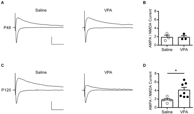
Figure 2.
AMPA—NMDA ratio is increased in adult but not adolescent VPA mPFC neurons. (A) Example EPSCs recorded from visually identified pyramidal neurons in layer V/VI mPFC from adolescent rats (P45–P49). Negative traces represent inward evoked currents from neurons patch-clamped at −70 mV, positive traces represent outward evoked currents from neurons patch-clamped at +40 mV. Scale bar 50 ms, 100 pA. (B) Average AMPA/NMDA ratio from adolescent rats exposed to saline or VPA in utero. (C) Similar evoked example EPSC traces from visually identified deep layer pyramidal neurons from adult rats (P110–P130). (D) Average AMPA/NMDA ratio from adult rats exposed to saline or VPA in utero. Data points represent AMPA/NMDA ratio from individual animals. Data shown as mean ± s.e.m.; * p < 0.05.

