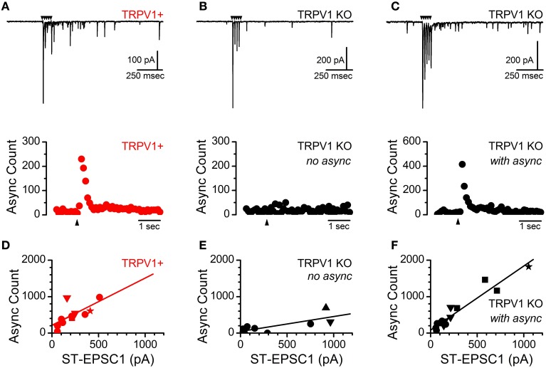
Figure 4.
Identification of a subpopulation of ST afferents from TRPV1 KO mice which maintain activity dependent asynchronous glutamate release. (A, upper) Current trace showing ST driven synchronous and asynchronous release from an identified TRPV1+ afferents in control mice. (A, lower) Histogram of summed asynchronous quantal glutamate events from across 50 trials. Black arrows indicate time of ST shock in all panels. (B, upper/lower) Nearly half (6 of 13) of all neurons recorded from TRPV1 KO ST afferents showed no asynchronous release regardless of the size or number of direct ST inputs. (C, upper) A significant population of TRPV1 KO afferents still mobilized vesicles via the asynchronous release pathway; with the magnitude and duration of additional release similar to the TRPV1+ afferents from control animals (C, lower). (D) The total number of asynchronously released vesicles positively correlates with the size of the evoked ST-EPSC (n = 11 inputs, R2 = 0.48, slope = 1.34 ± 0.47) from control TRPV1+ afferents. (E) TRPV1 KO afferents with no asynchronous release show only a weak relationship between EPSC1 amplitude and additional quantal events (n = 8 inputs, R2 = 0.58, slope = 0.42 ± 0.14). (F) From TRPV1 KO afferents with asynchronous release the magnitude of the release was strongly correlated with the size of the evoked synchronous EPSC (n = 15 inputs, R2 = 0.92, slope = 1.75 ± 0.14). The synchronous to asynchronous relationship is maintained across multiple inputs and the data have been fit including convergent ST contacts: 1st = circle, 2nd = down triangle, 3rd = square, 4th = star, and 5th = upward triangle. These data suggest an additional mechanism of mobilizing asynchronous glutamate vesicles is present in a subpopulation of ST afferents.

