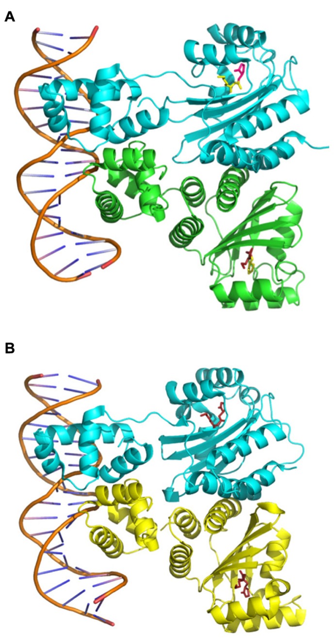
FIGURE 3.
Structures of the TraR–OC8HSL dimers in complex with DNA. The images were created using data from The Protein Data Bank (PDB; www.rcsb.org) (Berman et al., 2000) and the PyMOL Molecular Graphics System software. (A) PDB ID: 1H0M from Vannini et al. (2002). (B) PDB ID: 1L3L from Zhang et al. (2002b).

