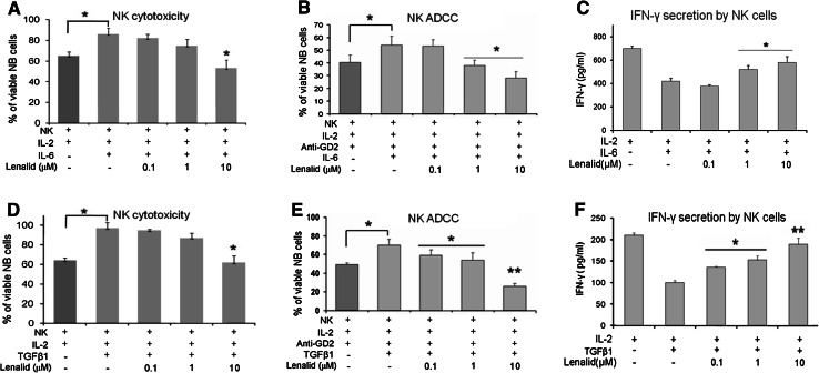
Fig. 3.
Suppression of IL-2 induction of NK cell direct cytotoxicity, ADCC, and IFNγ secretion by IL-6 and TGFβ1 is prevented by lenalidomide. a, b Purified NK cells (1 × 10⁴ cells/0.1 ml/well) were cultured for 72 h with IL-2 alone (10 ng/ml) or with added IL-6 (10 ng/ml) and lenalidomide as indicated, and then NK cell-mediated cytotoxicity and ADCC (E:T ratio = 2:1) with ch14.18 (0.1 μg/ml) were quantified after 6 h of co-culture with CHLA-255-Fluc cells with the calcein-AM/DIMSCAN assay (mean ± SD for 8 replicate cultures for each condition). Confirmatory results were obtained from 3 additional experiments. c NK cells (5 × 10⁵/ml) were cultured for 24 h with IL-2 alone (10 ng/ml) or with added IL-6 (10 ng/ml) and lenalidomide as indicated, and then IFNγ was quantified in the medium by ELISA (mean ± SD for 3 replicate cultures for each condition). Confirmatory results were obtained with 2 additional experiments. d, e NK cells (1 × 10⁴ cells/0.1 ml/well) were cultured for 72 h with IL-2 alone (10 ng/ml) or with added TGFβ1 (10 ng/ml) and lenalidomide as indicated, and then NK cell-mediated cytotoxicity and ADCC (E:T ratio = 2:1) with ch14.18 (0.1 μg/ml) were quantified after 6 h of co-culture with CHLA-255-Fluc cells with the calcein-AM/DIMSCAN assay (mean ± SD for 8 replicate cultures for each condition). Confirmatory results were obtained from 3 additional experiments. f NK cells (5 × 10⁵ cells/ml) were cultured for 24 h with IL-2 alone (10 ng/ml) or with added TGFβ1 (10 ng/ml) and lenalidomide as indicated, and then IFNγ was quantified in the medium by ELISA (mean ± SD for 3 replicate cultures for each condition). Confirmatory results were obtained with 1 additional experiment. The t test P values, no lenalidomide versus lenalidomide *P < 0.05; **P < 0.01

