Figure 6.
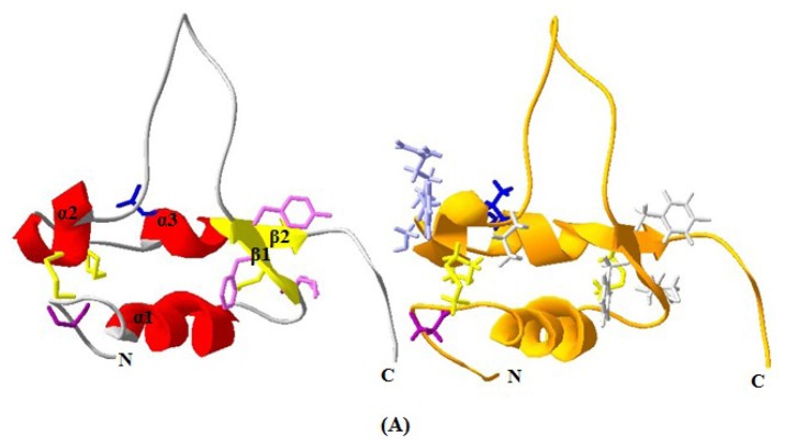
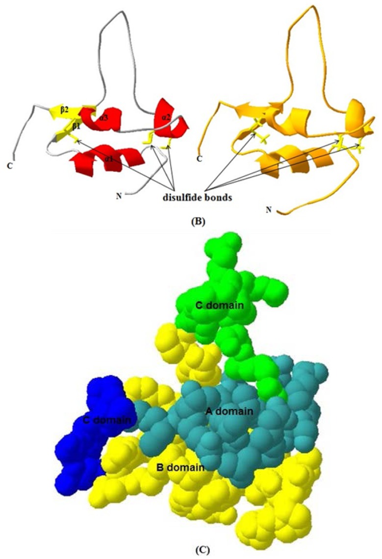
Predicted 3D structure model of yak IGF2 and the mainly chain interface structure. The 3D structure model was predicted by Swiss-model server. Left: yak IGF2, right: human IGF2. (A) Each chain contains 3 helixes. Binding sites were shown as rodlike molecules for yak IGFBP (modena), yak IGFBP and IGF-receptor and insulin-receptor (mauve) and yak IGF-receptor (blue); human IGFBP (modena), type 1 human IGF-receptor and insulin-receptor (white), type 2 human IGF receptor and IGFBP (French grey), human IGF-receptor (blue); (B) The view of IGF2 reveals three disulfide bonds (yellow, rod-like) which stabilize the fold; and (C) The mainly chain have four domain which B-domain (yellow), C-domain (green), A-domain (cyan) and D-domain (blue).


