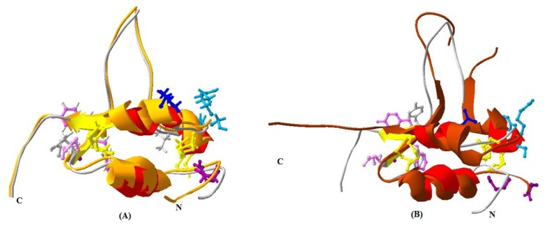
Figure 7.
Superimposed 3D structure: Tianzhu white yak IGF2 (red) with human IGF2 (Ginger) (A) and human IGF1 (brown) (B). Binding sites were shown as rodlike molecules. The superimposition indicated very high similarity between the structures of yak IGF2 and the 3D structure of human IGF2. Binding sites were shown as rodlike molecules for yak IGFBP (modena), yak IGF-receptor and insulin-receptor and IGFBP (mauve) and yak IGF-receptor (blue); human IGFBP (modena), type 1 human IGF-receptor and insulin-receptor (white), type 2 human IGF receptor and IGFBP (grey), human IGF-receptor (blue). The view of IGF2 revealed three disulfide bonds (yellow, rod-like) which stabilize the fold.

