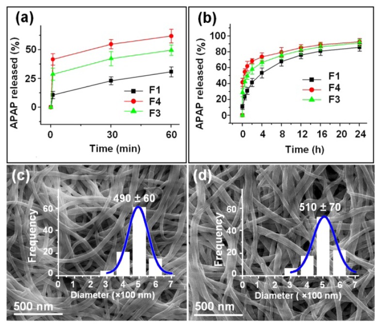
Figure 6.
In vitro dissolution tests: (a and b) In vitro drug release profiles of the APAP-loaded nanofibers F1 (prepared by single fluid electrospinning), F3, and F4 (prepared by coaxial electrospinning) at the first 60 min and the full range, respectively; (c and d) FESEM images and size distribution of the core part of nanofibers F3 and F4 after the removal of the shell part in the first phase.

