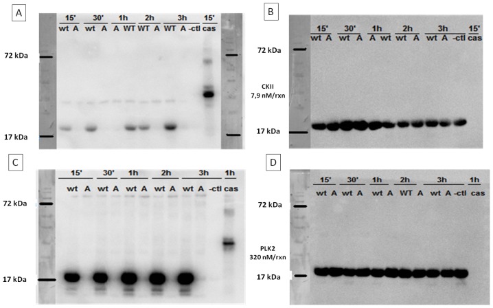
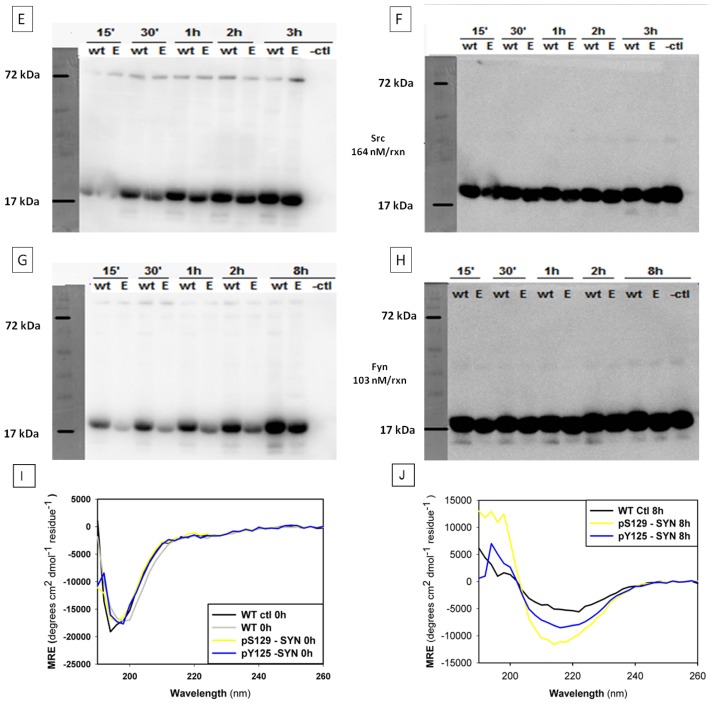
Figure 2.
Phosphate incorporation using autoradiography and far UV-CD spectra of (phosphorylated)-α-SYN. Phosphate incorporation was monitored by a radioactivity assay, where ATP-P32 was incubated in the presence of different kinases and WT α-SYN or the S129A/ Y125E mutant. The reaction was stopped at different time points (15 min, 30 min, 1 h, 2 h, 3 h as well as 24 h for PLK2 and Fyn) with SDS-loading dye and boiling for 5 min. Radioactive phosphate incorporation was visualised by autoradiography, after which western blotting was performed to detect relative protein levels. For each time point WT and a respective mutant were used. WT: WT α-SYN, A: S129A α-SYN, E: Y125E α-SYN, -ctl: negative control (kinase boiled for five min prior to test). Cas: casein kinase, used as a positive control for serine phosphorylation. Kinases and the kinase concentrations used in the in vitro phosphorylation assays are given between the blot panels (please refer to the Materials and Methods section for full details on the kinases and phosphorylation procedure) (A) radio-activity blot of CKII phosphorylation; (B) western blot of (A); (C) radioactivity blot of PLK2 phosphorylation; (D) western blot of (C); (E) radioactivity blot of SRC phosphorylation; (F) western blot of (E); (G) radioactivity blot of Fyn phosphorylation; (H) western blot of (G); (I) Far UV-CD spectra of proteins after overnight phosphorylation. All spectra show a minimum near 200 nm comparable to that of WT α-SYN (black line), typical for a random coil structure. The manipulations necessary for phosphorylation and removal of ATP afterwards do not disturb the secondary structure (compare spectrum of WT control (black line) to that of the WT protein (gray line)). pY125-α-SYN: WT α-SYN phosphorylated by Fyn kinase (blue line) and pS129-α-SYN: WT α-SYN phosphorylated by PLK2 kinase (yellow line); and (J) Far UV–CD spectra of pS129-α-SYN (yellow line), pY125-α-SYN (cyan) and their control (WT ctl (black line)), prepared as in (I), after 8 h of continuous agitation. All samples show predominantly a β-sheet structure.


