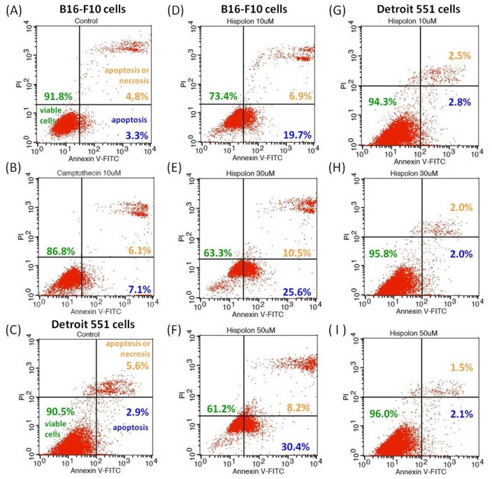
Figure 6.
Effects of hispolon on apoptosis in B16-F10 and Detroit 551 cells. The cells were stained with fluorescein isothiocyanate (FITC)-labeled annexin-V/propidium iodide (PI) double stain, and the percentages of apoptotic and necrotic cells were calculated. (A) untreated B16-F10 cells; (B) 10 μM camptothecin treated B16-F10 cells; (C) untreated Detroit 551 cells; (D) B16-F10 cells treated with 10 μM of hispolon; (E) B16-F10 cells treated with 30 μM of hispolon; (F) B16-F10 cells treated with 50 μM of hispolon, (G) Detroit 551 cells treated with 10 μM of hispolon; (H) Detroit 551 cells treated with 30 μM of hispolon; and (I) Detroit 551 cells treated with 50 μM of hispolon.

