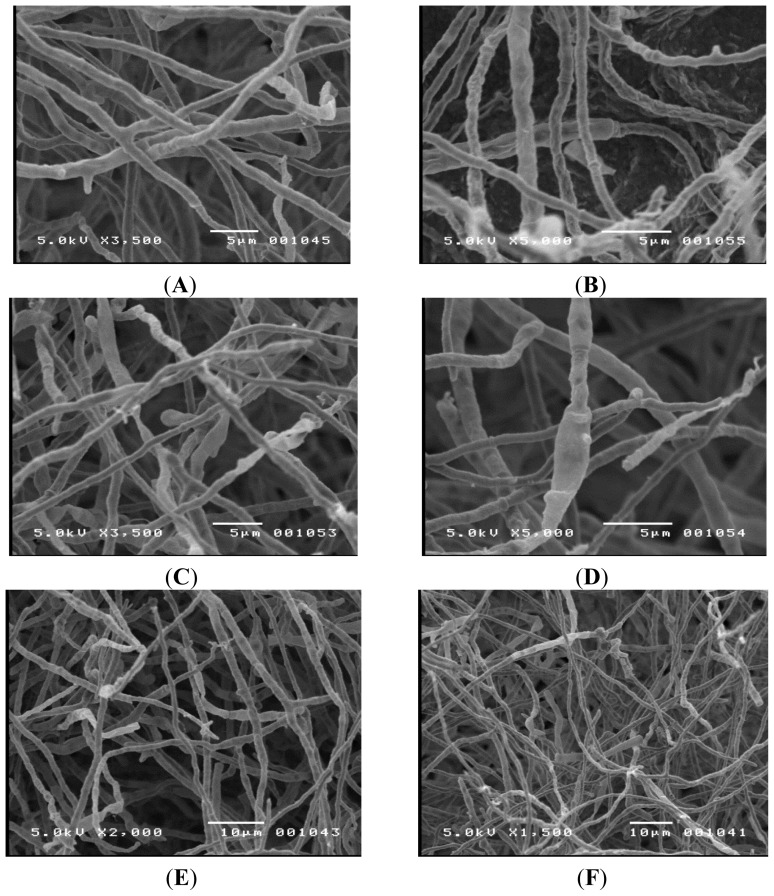
Figure 2.
Scanning electron microscopy images of TM-417 performed on a JEOL JSM-5400 scanning electron microscope (JEOL; Tokyo, Japan). No identifying structures were noticed. (A) 3500× magnification; (B) 5000× magnification; (C) 3500× magnification; (D) 5000× magnification; (E) 2000× magnification; (F) 1500× magnification. A swollen cell can be seen in (D).

