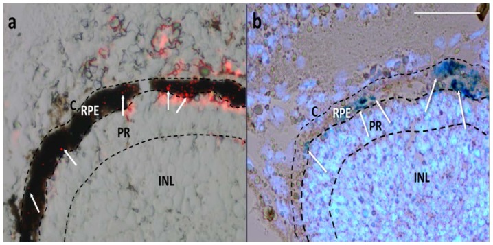
Figure 2.
Cryostat sections of Xenopus embryos 1 day after injection. (a) Merged image of bright field (showing the pigmented RPE) and red fluorescence field (showing MNPs as red spots); (b) Image from a bleached section merging the bright field (showing MNPs as dark blue spots by Prussian Blue staining) and fluorescence field (showing fluorescent blue nuclei by Hoechst staining). White arrows point some MNPs. C, choroid; RPE, retinal pigmented epithelium; PR, photoreceptors; INL, inner nuclear cell layer. Scale bar, 50 μm.

