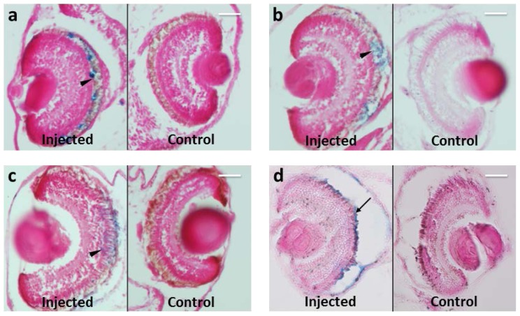
Figure 4.
Prussian Blue staining on paraffin section of Xenopus embryos at different time points from MNPs injection. MNPs are blue labeled. n = 45 each time point. (a) Three days after injection; (b) five days after injection; (c) 10 days after injection; (d) 20 days after injection. Arrowheads point to MNP localization in interdigitated RPE microvilli with the outer segment of photoreceptors; arrow points to MNPs derived from RPE rupture, during manipulation (details in the material and methods section). Scale bar, 50 μm.

