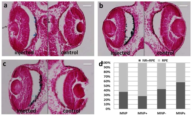
Figure 6.
Prussian Blue staining on paraffin section of Xenopus embryos one day after injection. Particles are blue labeled. n = 45 each group. (a) Left eye injected with MNP−; (b) Left eye injected with MNP+; (c) Left eye injected with MNPs; (d) Graphical representation of MNP, MNP+, MNP− and MNPs localization in eye regions of the embryo population. VC, vitreous chamber; NR, neural retina; RPE, retinal pigmented epithelium. Scale bar, 50 μm.

