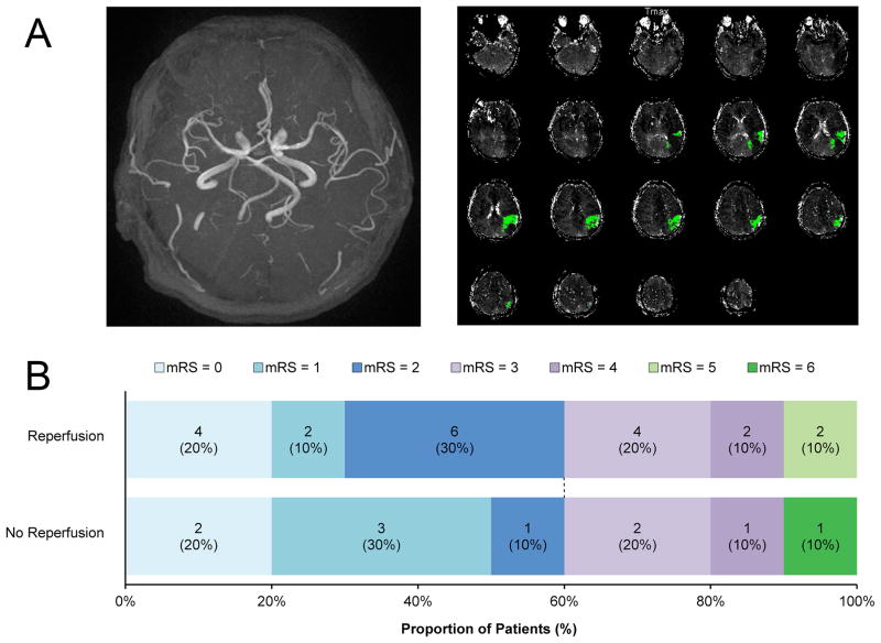
Figure 1. Example of imaging and functional outcome according to mRS at 90 days.
A An example of a patient included in the pooled analysis. MRA did not reveal vessel occlusion (left panel), but a single distal MCA perfusion lesion (right panel) was visualized on PWI (Tmax>6 sec).
B Good functional outcome (score 0–2) did not differ between patients in relation to reperfusion (p=1.0). Defining outcome more strictly as an mRS of 0–1 (excellent outcome) did not alter the results (30% of patients with reperfusion versus 50% of patients without reperfusion; p=0.4)
mRS= modified Rankin scale.

