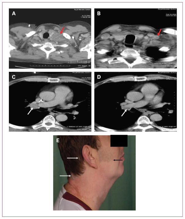
Fig. 2.
Pretreatment and posttreatment computed tomography imaging data for target (A and B) and nontarget (C and D) lesions of patient 0101. A and B, pretreatment and posttreatment imaging of left supraclavicular fossa mass (red arrows) showing significant volume reduction maintained at 7 months (B). C, D, pre- and posttreatment imaging of mediastinal disease (white arrows) showing a maintained response at 7 months (D). E, Vitiligo arising in the irradiated field of patient 0602. Note the sharp demarcation between the normal pigmentation in the hair-bearing skin and the depigmented, nonhearing bearing skin at the superior border of the radiation field (black arrow). Focal loss of hair pigmentation is also seen (white arrows).

