
Figure 2.
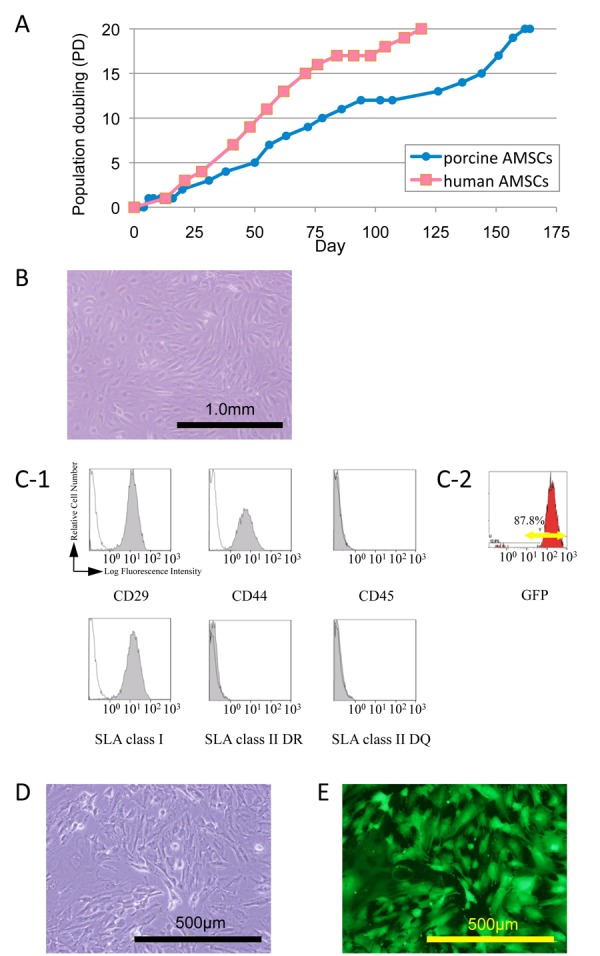
Characterization of porcine amniotic membrane-derived mesenchymal stromal cells (AMSCs). (A) The growth curves of porcine AMSCs used in this experiment compared to human AMSCs from a previous paper (ref. 28). (B) Phase contrast microscopic view of porcine AMSCs. (C-1) Flow cytometric analysis of porcine AMSCs using antibodies for CD29, CD44, CD45, swine leukocyte antigen (SLA) class I, SLA class II DR, and SLA class II DQ. Black lines and shaded areas indicate reactivity of antibodies for isotype controls and that of antibodies for cell surface markers, respectively; (C-2) Representative flow cytometrical GFPexpressing pattern in GFP-expressing porcine AMSCs. (D, E) Microscopic view of porcine GFP-expressing AMSCs.