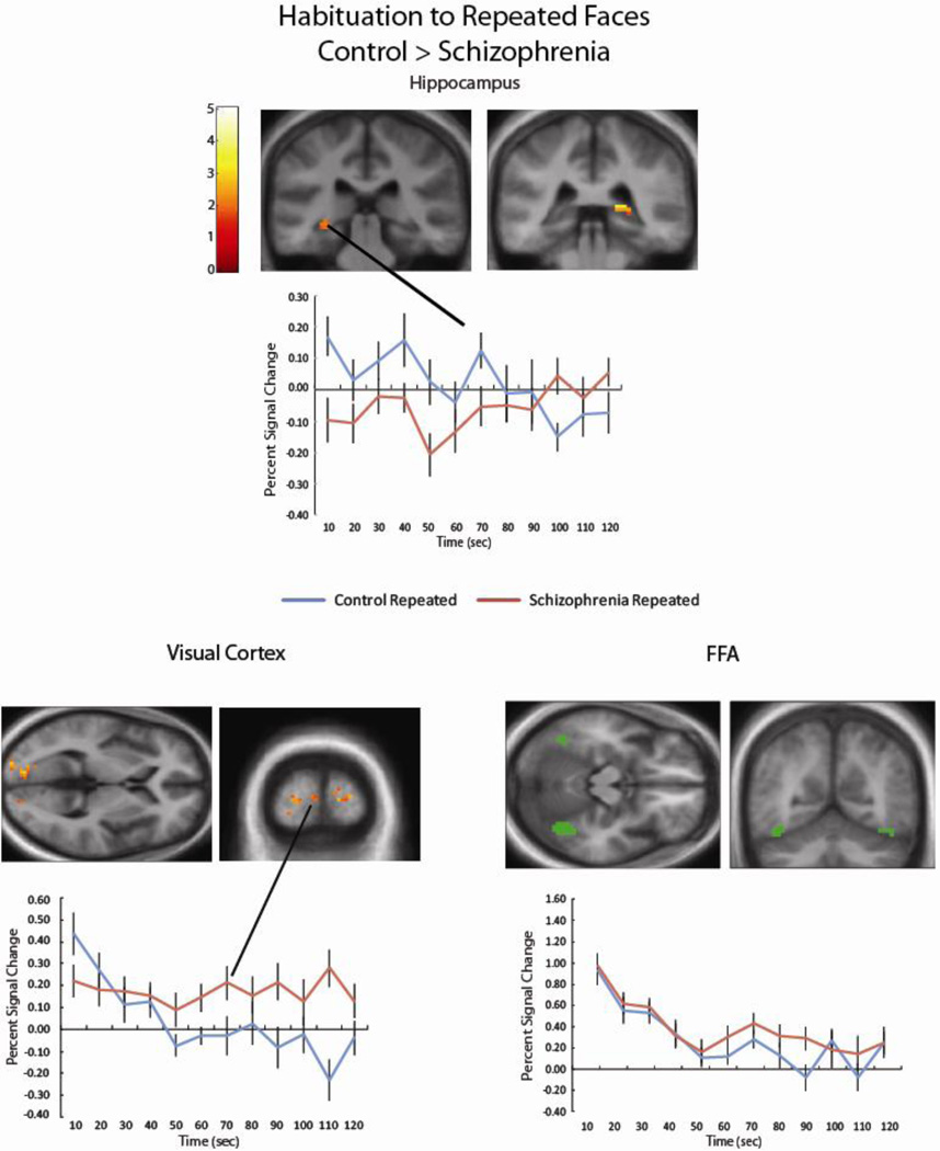
Figure 2.
Patients with schizophrenia showed reduced habituation to Repeated faces in the left (k = 34; − 21, −22, −23) and right (k = 13; 18, −37, 4) hippocampus (cluster corrected p-value < .05). Extracting the percent signal change from these clusters shows that while control participants show decreasing hippocampal activity across the run, activity is more sustained for schizophrenia patients. A similar pattern was found in three clusters in the bilateral visual cortex (k = 111, −18, −79, 10; k = 22, 12, −76, 22; k = 19, 24, −97, 7, cluster-corrected p < .05). Percent signal change showed for a representative cluster. In contrast, both controls and schizophrenia patients show robust habituation to Repeated faces within the FFA (a representative single-subject FFA region of interest shown in green).

