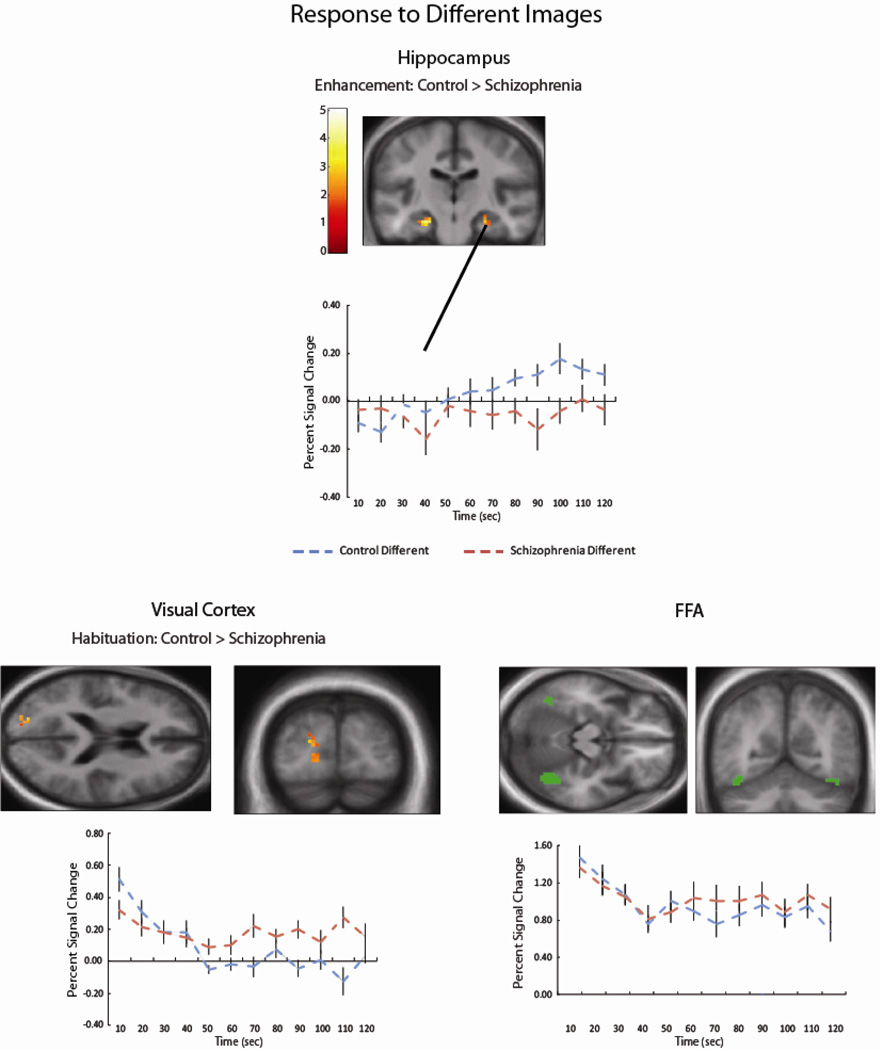
Figure 3.
In response to Different Images, control subjects showed increasing signal in the bilateral hippocampus (left k = 56, −27, −40, 1, k = 14, −24, −19, −26; right k = 27, 33, −37, −8, k = 17, 27, −22. −26 cluster-corrected p < .05); schizophrenia patients did not show significant hippocampal changes throughout the run. In the visual cortex, patients with schizophrenia showed reduced habituation to Different faces, relative to control participants (k = 53, −21, −88, 16 cluster-corrected p < .05). FFA habituation to Different faces was present for both groups.

