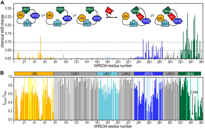
FIGURE 5.
Chemical shift perturbation mapping of Vpr binding to full-length hHR23A and PRE data on free full-length hHR23A. A, 1H,15N-combined chemical shift changes (calculated using the formula given in the legend for Fig. 3B) in the 1H-15N HSQC spectrum of hHR23A upon Vpr binding. Inset, schematic of intramolecular and intermolecular interactions in hHR23A upon Vpr binding. B, PRE effects on individual resonances were estimated by calculating the resonance intensity ratio from 1H-15N HSQC spectra of hHR23A with (Ipara) and without (Idia) MTSL labeling at Cys-344. Sample conditions were identical for both. Chemical shift changes (A) and PRE effects (B) are color-coded according to domain identity: UBL (orange); LNK1, LNK2, and LNK3 (gray); UBA1 (light blue); XPCB (blue); and UBA2 (green). PRE values for prolines and unassigned residues were arbitrarily set to 1 and colored according to domains using a lighter color scheme.

