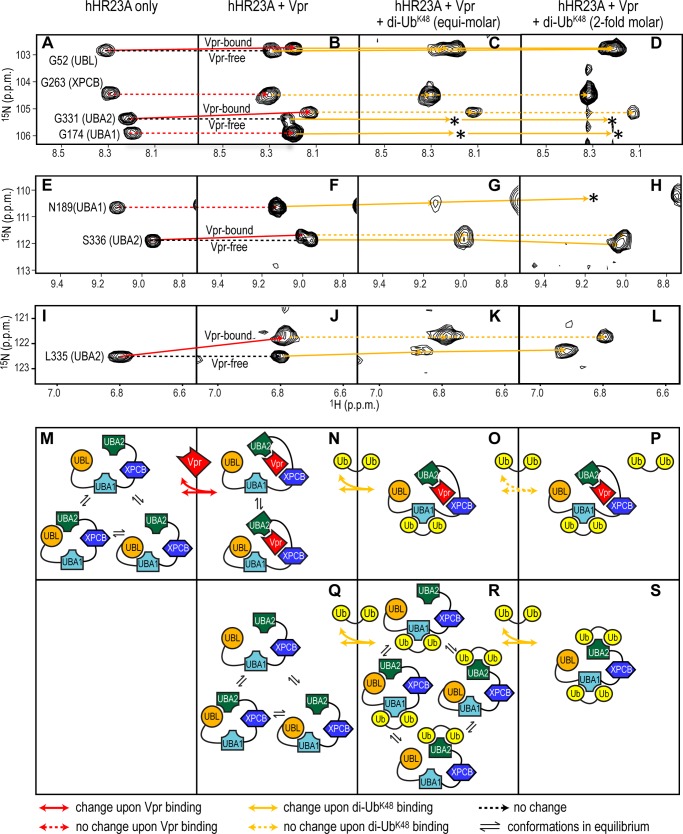
FIGURE 6.
Vpr and di-UbK48 binding to hHR23A. Selected 1H-15N HSQC resonances, representative of the UBL, UBA1, XPCB, and UBA2 domains in free hHR23A (A, E, and I), hHR23A-Vpr (B, F, and J) and hHR23A-Vpr complex samples with equimolar (C, G, and K) or 2-fold excess (D, H, and L) di-UbK48, were monitored and connected by solid (chemical shift change) or dashed (no chemical shift change) lines. Red and orange are used for Vpr- and di-UbK48-induced changes, respectively. Loss of resonances because of extensive broadening or other causes are indicated by asterisks. M–S, schematics summarizing the intramolecular and intermolecular interactions.

