FIGURE 1.
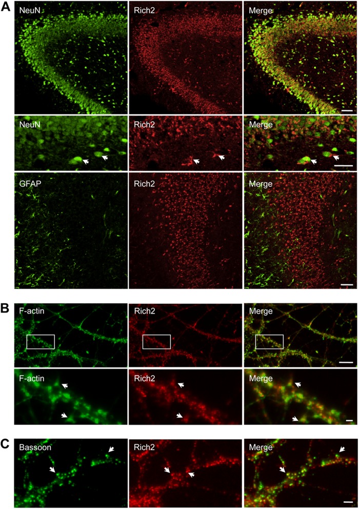
Rich2 is localized in dendritic spines. A, immunostaining of E18.5 rat hippocampal CA1, CA2, and CA3 regions with anti-NeuN (green) and anti-Rich2 (red) antibodies (upper panel). In this and the following panels, right images represent merged red and green images. Middle panel: similar to the upper panel, but in an enlarged region of CA1 of a different sample. Lower panel: immunostaining of E18.5 rat hippocampal with an anti-GFAP antibody. Please note localization of Rich2 in neurons (NeuN labeling, arrows), but not glial cells (GFAP labeling; Scale bars: 100 and 40 μm, respectively). B, DIV-21 hippocampal neurons treated with phalloidin (green) and anti-Rich2 antibody (red) to visualize F-actin and endogenous Rich2, respectively. Arrows indicate co-localization of Rich2 and actin in dendritic spines (scale bars: 10 and 1 μm, respectively). C, dendritic portion of a DIV-15 cultured hippocampal neuron immunolabeled with an anti-bassoon antibody showing the synaptic localization of Rich2, (arrows, scale bar: 1 μm).

