
FIGURE 2.
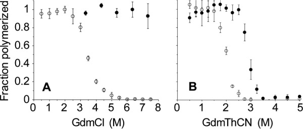
rPrP amyloid fibrils formed in 2 and 4 m GdmCl have different conformational stabilities. A, denaturation profiles in GdmCl. B, denaturation profiles in GdmThCN. In each case, the open and closed circles represent fibrils formed in 2 and 4 m GdmCl, respectively.