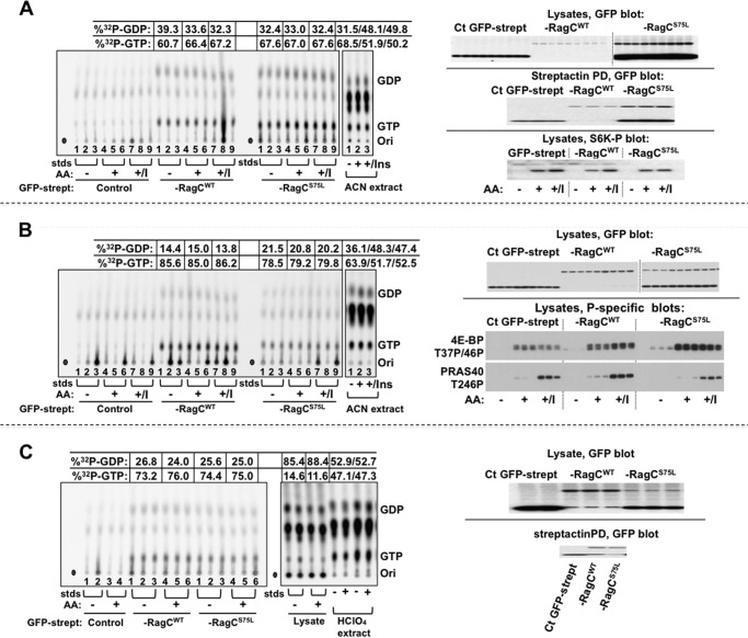
FIGURE 6.
The effect of amino acid withdrawal and insulin on the [32P]guanyl nucleotide content of stably expressed RagCWT and RagCS75L heterodimeric complexes in 32Pi-labeled HeLa cells. A, B, and C, replicate plates of HeLa cells stably expressing GFP-streptag, GFP-streptag-RagCWT, or GFP-streptag-RagCS75L were incubated in Pi-free DMEM containing 32Pi (0.2 mCi/ml). After 4 h, the cells were rinsed and incubated in either homemade Pi-free medium (AA+) or Pi-free medium lacking amino acids (AA-), each containing 32Pi (0.2 mCi/ml), for another 2 h. In A and B, insulin (1.0 μm) was added to some of the cells in DMEM (AA+/I) 30 min before harvest. The nucleotides bound to Strep-Tactin (strept) pull-downs were extracted and separated by TLC on PEI cellulose. One set of 32P -labeled HeLa cells expressing FP-streptag, treated as described above for AA-, AA+, and AA+/I, were rinsed, extracted directly into acetonitrile, and then the solubilized total nucleotides were separated by TLC. The 32P comigrating with GDP and GTP was quantitated by phosphorimaging. After subtraction of the averaged values found in the GFP-streptag lanes, the percentage of [32P]GTP was calculated as [[32P]GTP / (1.5 × [32P]GDP + [32P]GTP)], and the averaged values are shown. In A, the top and center panels on the right show GFP immunoblot analyses of the cell lysates and representative Strep-Tactin pull-downs (corresponding to lanes 1, 4, and 7 of each set). The bottom panel shows an immunoblot analysis of S6K(T389P) corresponding to lanes 1, 4, and 7 of each set. In B, the top panel on the right shows a GFP immunoblot analysis of the lysates, whereas the center and bottom panels show lysate immunoblot analyses for 4E-BP(T37P/T46P) and PRAS40(S246P), respectively. In C, one set of 32P-labeled HeLa cells expressing GFP-streptag, treated as in Fig. 6, were rinsed, extracted directly into HClO4 (0.3 m, 0 °C, HClO4 extract). The HClO4 supernatants were neutralized with KHCO3, and the [32P]guanyl nucleotides were quantified as above. In addition, the nucleotides in the lysate after pull-down of the Strep-Tactin beads were also analyzed. Immunoblot analyses of the lysates and representative Strep-Tactin pull-downs are shown in the right panel. Ori, origin; std, guanyl nucleotide standards; Ins, insulin; ACN, acetonitrile; Ct, C-terminal.

