FIGURE 2.
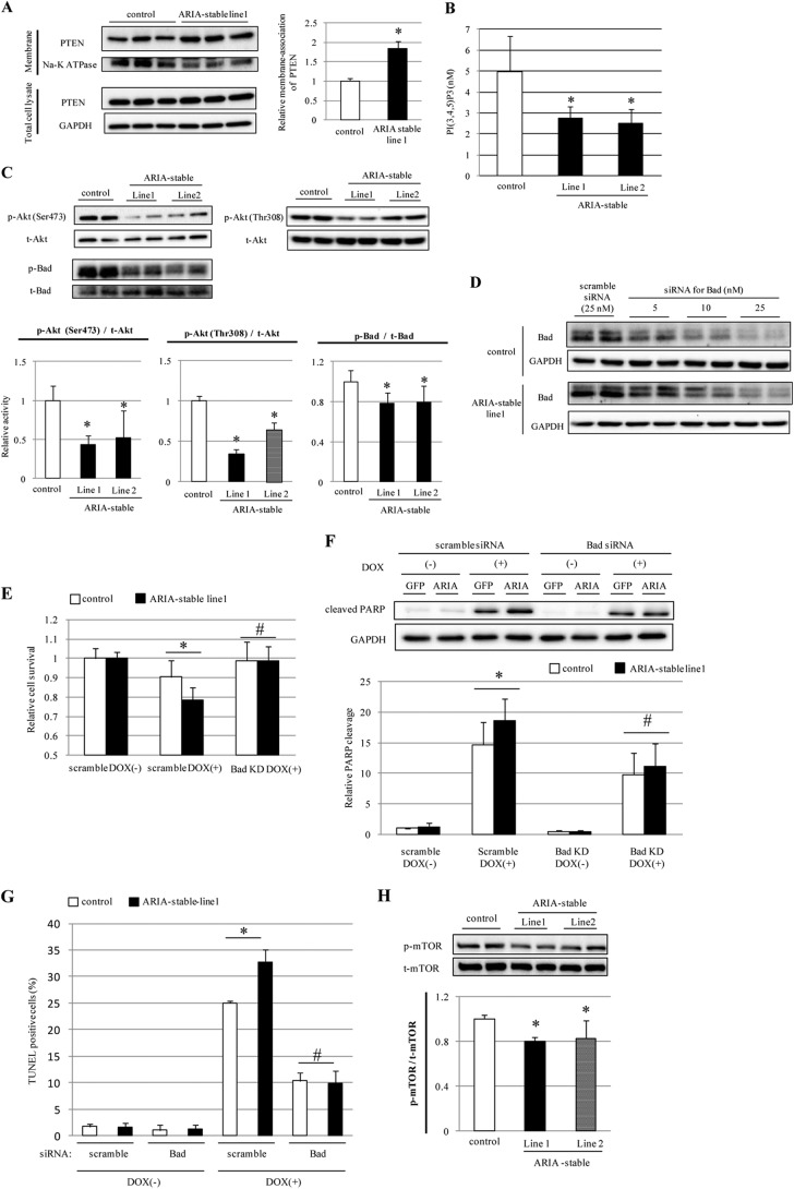
ARIA regulates the cardiomyocyte apoptosis through a modification of the PI3K/Akt/Bad axis. A, expression of PTEN in the membrane fraction of H9c2 cells stably expressing GFP (control) or ARIA (ARIA-stable) (n = 6). *, p < 0.05 versus control. B, phosphatidylinositol 1,4,5-trisphosphate contents in lipid extracts of H9c2 cells stably expressing GFP (control) or ARIA (ARIA-stable) (n = 4). *, p < 0.05 versus control. C, phosphorylation of Akt and Bad was assessed by immunoblotting in H9c2 cells stably expressing GFP (control) or ARIA (ARIA-stable) (n = 6). *, p < 0.05 versus control. D, H9c2 cells stably expressing GFP (control) or ARIA (ARIA-stable) were transfected with either scramble or Bad siRNA at the indicated concentrations. E, cell survival was assessed by MTS assay in H9c2 cells stably expressing GFP (control) or ARIA (ARIA-stable) (n = 8). Cells were transfected with either scramble or Bad siRNA (Bad KD). *, p < 0.05; #, not significant versus control. F, DOX (1 μm)-induced apoptosis was analyzed by detecting the poly(ADP-ribose) polymerase (PARP) cleavage (n = 6). Cells were transfected with either scramble or Bad siRNA (Bad KD). *, p < 0.05; #, not significant versus control. G, DOX (1 μm)-induced apoptosis was analyzed by TUNEL staining (n = 3). Cells were transfected with either scramble or Bad siRNA. *, p < 0.05; #, not significant versus control. H, phosphorylation of mTOR was assessed by immunoblotting in H9c2 cells stably expressing GFP (control) or ARIA (ARIA-stable) (n = 5). *, p < 0.05 versus control.

