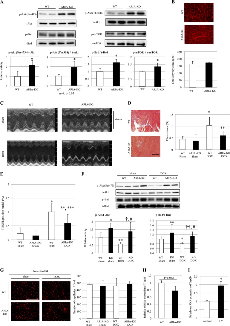
FIGURE 4.
ARIA regulates the cardiac PI3K/Akt signaling and modifies the progression of DOX-induced cardiomyopathy. A, phosphorylation of Akt, Bad, and mTOR in the hearts of WT or ARIA-KO mice (n = 5). *, p < 0.05 versus WT mice. B, cardiomyocyte size was evaluated by staining the heart sections with wheat germ agglutinin (n = 5). Bar = 50 μm. C, representative M-mode images of echocardiograms in sham- or DOX-treated WT or ARIA-KO mice. D, cardiac fibrosis was evaluated by the Masson's trichrome staining (n = 6). *, p < 0.05 versus sham-treated WT mice. **, p < 0.01 versus DOX-treated WT mice. Bar = 100 μm. E, cardiomyocyte apoptosis was evaluated by TUNEL staining (n = 5). *, p < 0.01 versus sham-treated WT mice. **, p < 0.01 versus sham-treated KO mice. ***, p < 0.05 versus DOX-treated WT mice. F, phosphorylation of Akt and Bad in the hearts of WT or ARIA-KO mice on either sham or DOX treatment (n = 6). *, p < 0.01 versus sham-treated WT mice. **, p < 0.05 versus sham-treated WT mice. †, p < 0.01 versus DOX-treated WT mice. ††, p < 0.05 versus DOX-treated WT mice. #, not significant versus sham-treated KO mice. G, capillary density was assessed by isolectin IB4 staining of the heart sections (n = 4). Bar = 100 μm. H, expression of Top2b mRNA was assessed by quantitative PCR in the hearts of WT or ARIA-KO mice (n = 4; p = 0.062 versus WT mice). I, expression of Top2b mRNA was assessed by quantitative PCR in rat neonatal cardiomyocytes treated with either vehicle (control) or PI3K inhibitor LY294002 (LY). n = 4. *, p < 0.05 versus control.

