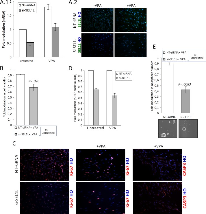
FIGURE 6.
Effects of si-SEL1L and VPA combined treatments in G179. A, 1 and 2, SEL1L mRNA and protein silencing efficiency after VPA treatment. G179 cell line (1 × 10⁶) was nucleofected with NT siRNA or two si-SEL1L for 48 h and maintained in the absence or presence of VPA for an additional 4 days. The data reflect the average of two si-SEL1L and two independent knockdown experiments. Silencing efficiency was verified by mRNA using qPCR (1) and protein using immunofluorescent staining (2). 1, the histogram shows values normalized relative to housekeeping signals and are expressed as -fold modulation relative to controls. HO, Hoechst. B–E, the G179 was treated with NT siRNA or si-SEL1L for 48 h and analyzed as follows. B, MTT cell viability assay. Nucleofected cells were seeded in 96-well plates at a density of 2000. After 24 h, cells were treated with VPA and analyzed for viability. The histogram shows cell viability values expressed as -fold modulation relative to drug-treated (VPA) versus untreated samples. p was used to determine statistical significance. C, Immunofluorescence analysis. Nucleofected cells were seeded on 12-well cover glass and maintained in the absence or presence of VPA for 4 days. A decrease in the number of positive Ki-67 cells was observed in si-SEL1L G179s with respect to control either in the absence or presence of VPA. Scattered areas with faint CASP3 immunoreactivity were observed in treated si-SEL1L G179 cells. D, Ki-67 was evaluated in terms of % of positively stained nuclei counted in at least 800 cells per group. E, Neurosphere assay. Nucleofected cells were seeded in 6 low attachment plates wells at density of 3000 and maintained in neurosphere cell conditions in the absence or presence of VPA for 4 days. Then the culture was allowed to recover from drug treatment by adding fresh medium in excess for an additional 7 days. The figure is a representative image of three independent experiment. p was is used to determine statistical significance.

