
FIGURE 3.
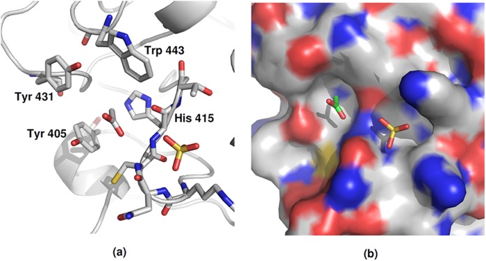
Acetyl binding site S1 in each protomer of the subunit A tetramer of the native FIBCD1 structure. The acetate and sulfate ions located in and in proximity to the S1 acetyl binding pocket are shown. a, key interacting amino acids. b, charged surface representation of the extended S1 site including the acetyl binding pocket and the adjacent pocket which accommodates a sulfate ion.