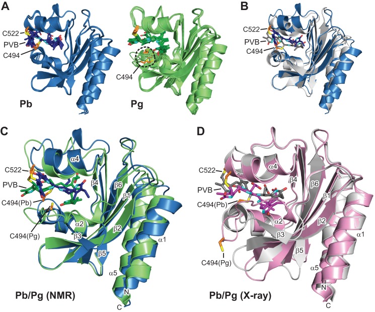
FIGURE 1.
Three-dimensional structures of the GAF domain from TePixJ in the Pb and Pg states. A, ribbon diagrams of the five lowest energy conformers for Pb (blue) and Pg (green) as determined by solution NMR spectroscopy. The relatively disordered N and C termini were truncated for clarity. B, superposition of the Pb crystal structure (gray) with its lowest energy solution structure (blue). C, superposition of the lowest energy solution structures for Pg (blue) and Pg (green). D, superposition of the crystal structures for Pb (gray) and Pg (pink). The positions of Cys-494 and Cys-522 that form thioether linkages to PVB are indicated. Cyan, pyrrole nitrogens; red, oxygens; orange and yellow, side-chain carbon and sulfur atoms of Cys-494 and Cys-522, respectively. The α-helices and β-strands are labeled as are the N terminus (N) and C terminus (C).

