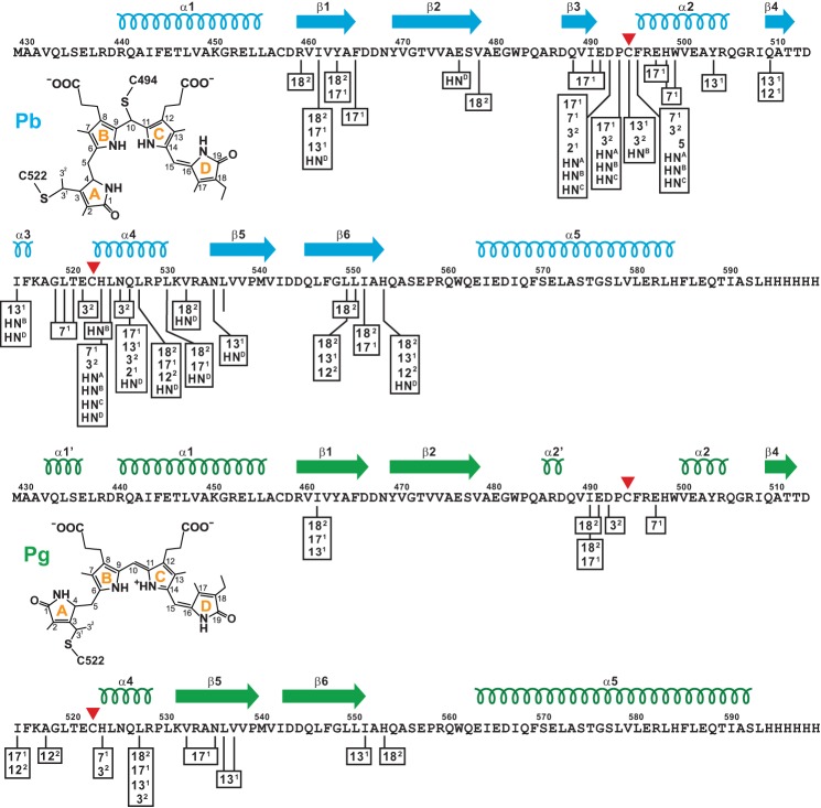
FIGURE 2.
Schematic diagram showing intermolecular NOE partners between the TePixJ(GAF) polypeptide (magenta) and the isotopically labeled 15N- or 13C-PVB as Pb (top, blue) and Pg (bottom, green). Cys-494 and Cys-522 that participate in the thioether linkages between the chromophore and polypeptide are located by the red arrowheads. The C-terminal His6 tag was included in the sequence. The locations of the predicted β-strands and α-helices are shown above the sequence. The proposed structures of bound PVB and carbon atom assignments for each state are included for reference. HN represents the pyrrole nitrogen protons (A-D rings are identified in superscript).

