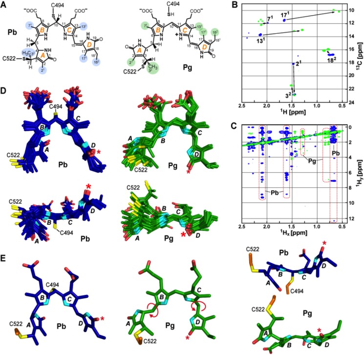
FIGURE 3.
Conformational rearrangements of PVB during TePixJ(GAF) photoconversion. A, proposed chemical structures of PVB bound to the GAF domain in the Pb and Pg states. The A-D pyrrole rings and the various pyrrole carbon atoms are labeled. Carbons highlighted in blue and green indicate the residues interrogated by NMR experiments for Pb and Pg, respectively. B, two-dimensional 13C,1H HSQC spectrum of TePixJ(GAF) with 13C incorporated into PVB carbons 21, 32, 71, 82, 122, 131, 171, and 182 as Pb (blue) and Pg (green). C, two-dimensional NOE spectrum of the sample in B showing the 1Hx/1Hy cross-peaks for Pb and Pg. Significant chemical shift changes for the 131, 171, and 182 carbons are highlighted. D, top and side views of the 10 lowest energy structures of PVB in the Pb (blue) and Pg (green) states as determined by solution NMR spectroscopy. E, top and side views of the lowest energy structures of PVB shown in panel D in the Pb (blue) and Pg (green) states. Light-induced rotation at the C5 and C15 bridges and the D ring flip are highlighted (the asterisk marks C17 carbonyl). Cyan, pyrrole nitrogens; red, oxygens; orange and yellow, side-chain carbon and sulfur atoms of Cys-494 and Cys-522, respectively.

