
FIGURE 4.
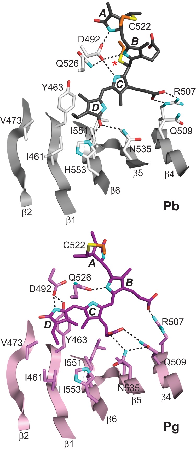
Repositioning of PVB in the TePixJ(GAF) pocket during photoconversion. The three-dimensional structures of the Pb (gray) and Pg states (pink) were determined by x-ray crystallography (21, 22). The relevant β-strands and amino acids are shown along with key hydrogen bond contacts (dashed lines). Cys-494 is located by an asterisk. Cyan, nitrogens; red, oxygens; orange and yellow, side-chain carbon and sulfur atoms of Cys-494 and Cys-522, respectively.