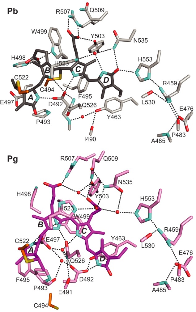
FIGURE 5.

Changes in bilin/protein contacts during Pb → Pg photoconversion of TePixJ(GAF). PVB is colored in gray and purple for the Pb (top) and Pg states (bottom), respectively. The A-D pyrrole rings and key amino acid contacts are labeled. Predicted hydrogen bonds are indicated by the dashed lines. Shown are the x-ray crystallographic structures (21, 22). Cyan, nitrogens; red, oxygens; orange and yellow, side-chain carbon and sulfur atoms of Cys-494 and Cys-522, respectively.
