
FIGURE 6.
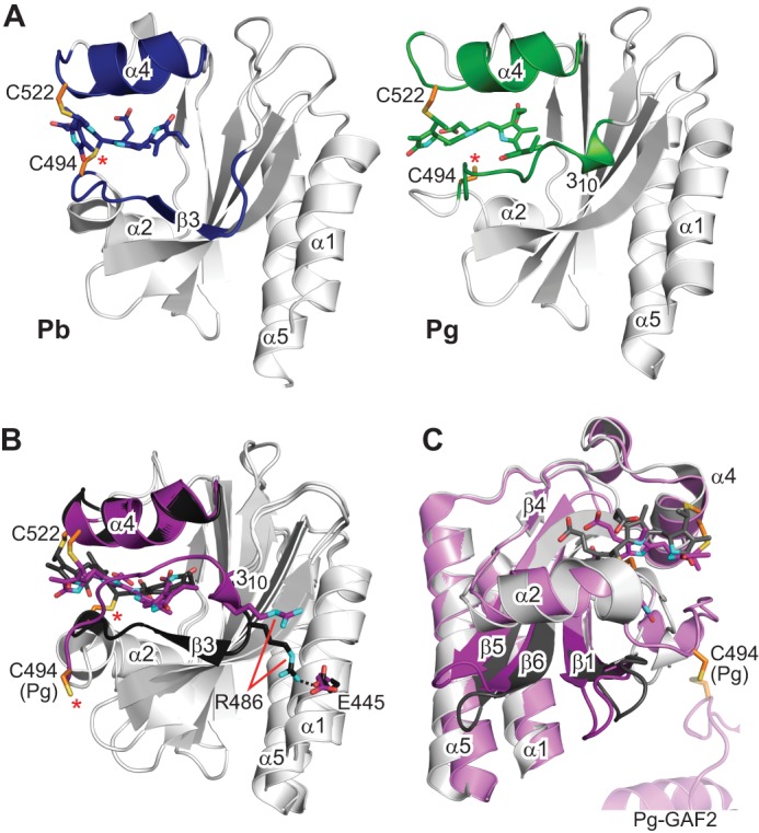
Large light-driven structural changes in TePixJ(GAF) influence the positions of helices α1 and α5. A, strand β3 is well defined in the solution NMR structure of Pb, but it dissolves into a dynamic loop region in Pg that is bounded by a 310-helix and helix α2. The position of the 310-helix draws this entire loop region closer to helix α4. B, the same structural perturbations are identified by comparison of the crystal structures of Pb and Pg (21, 22). Additionally, Pb → Pg photoconversion is coincident with rupture of the Arg-486–Glu-445 salt bridge, which may serve to loosen constraints on the helix α1 position. C, the alternative view provided by the paired crystallographic structures illustrates the sweeping movements of the loops between strands β1-β2 and β5-β6 and correlated motions of helices α1 and α5. The ectopic cystine disulfide found in the Pg crystal structure is included in C, with a portion of the second GAF domain from the asymmetric unit. Cys-494 is located by an asterisk. Cyan, nitrogens; red, oxygens; orange and yellow, side-chain carbon and sulfur atoms of Cys-494 and Cys-522, respectively.