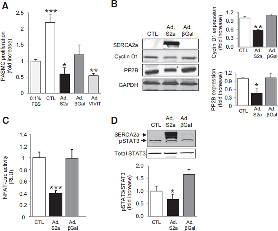
Figure 2.
SERCA2a gene transfer inhibits the proliferation of PASMCs. PASMCs were infected for 48 hours with Ad.SERCA2a, Ad.βGal, or Ad.VIVIT and then cultured for 48 hours in virus-free medium. A, Proliferation was determined by labeling cells with BrdU and supplementing the medium with 5% FBS to stimulate proliferation. Noninfected PASMCs and PASMCs cultured with 0.1% FBS to maintain quiescence cultured under the same conditions served as controls (n=6) *P=0.03 vs CTL. **P<0.005 vs CTL. ***P<0.001 vs CTL or 0.1% FBS. B, Western immunoblotting was performed to evaluate SERCA2a, protein phosphatase 2B (PP2B), and Cyclin D1 expression (n=3). Protein expression was normalized to GAPDH. *P<0.03 vs CTL. **P<0.008 vs CTL. C, NFAT transcriptional activity was assessed by using a luciferase promoter-reporter assay. Data are expressed in relative luciferase units (RLU; n=5) ***P<0.001 vs CTL. D, Phosphorylated and total STAT3 expression were determined by immunoblotting. Phosphorylated STAT3 was normalized to total STAT3. GAPDH was used as the loading control (n=3). Representative blots are shown. *P=0.04 vs CTL. BrdU indicates bromodeoxyuridine; CTL, control; FBS, fetal bovine serum; NFAT, nuclear factor of activated T cells; and PASMC, pulmonary artery smooth muscle cell.

