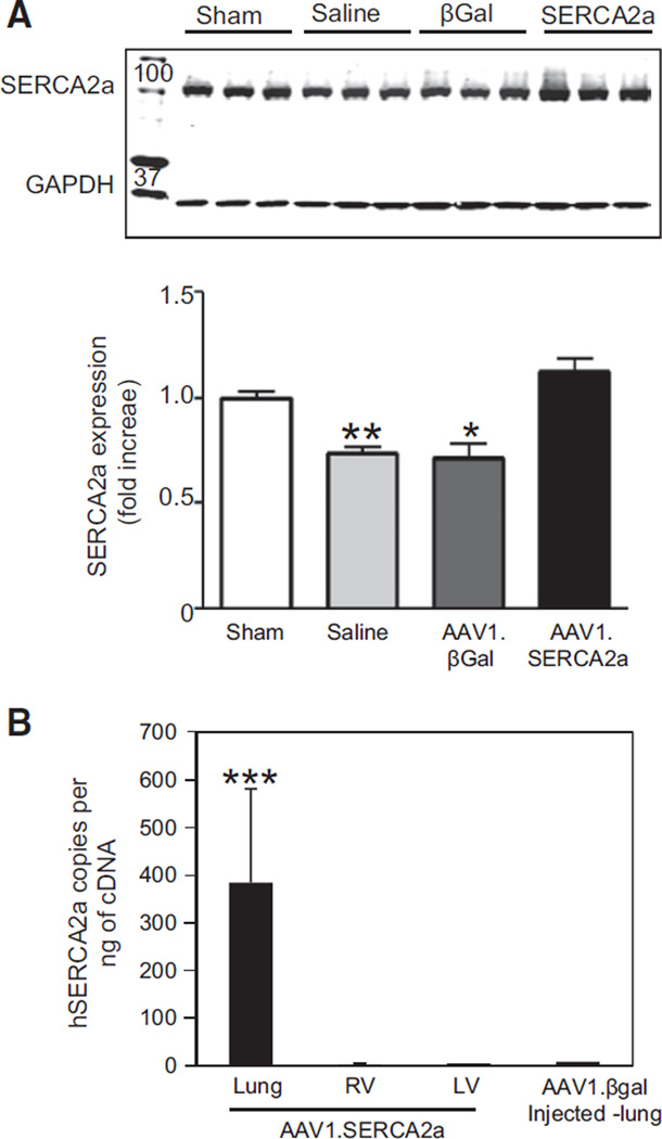
Figure 6.
SERCA2a expression in the RV in PAH. A, The RV was harvested from sham (n=6) and MCT-PAH rats treated with aerosolized saline (n=5), AAV1.βGal (n=5), or AAV1.SERCA2a (n=7), and homogenates were used to examine SERCA2a expression by Western blotting. A representative blot is shown (n=3). *P<0.02 vs sham, AAV1.SERCA2a, **P=0.003 vs sham, AAV1.SERCA2a. B, Levels of human SERCA2a and viral genome copies were assessed in the rat RV, LV, and lungs to determine the specificity of AAV1.SERCA2a gene transfer to lungs with aerosol delivery (n=3). ***P<0.001 vs RV, LV, and AAV1.βGal-injected lung. LV indicates left ventricle; MCT, monocrotaline; PAH, pulmonary arterial hypertension; and RV, right ventricle.

