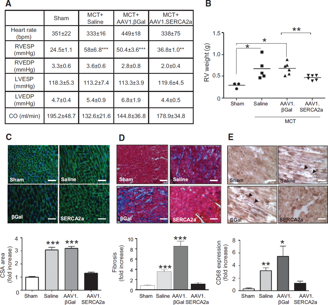
Figure 7.
AAV1.SERCA2a improves RV hemodynamics and decreases RV hypertrophy and fibrosis. A, Cardiac hemodynamics were measured invasively in sham (n=6) and MCT-PAH rats treated with aerosolized saline (n=5), AAV1.βGal (n=5), or AAV1.SERCA2a (n=7) at day 45. ***P<0.001 vs sham, AAV1.SERCA2a. **P<0.005 vs AAV1.βGal. B, RV hypertrophy was determined by the RV/LV+septum weight ratio (Fulton index). *P<0.04 vs sham, AAV1.SERCA2a, **P=0.006 vs AAV1.βGal. C, RV sections were stained with fluorescence-tagged wheat germ agglutinin to examine RV cardiomyocyte cross-sectional area (CSA; n=3 per animal). ***P<0.0001 vs sham, AAV1.SERCA2a. D, Collagen deposition and fibrosis was examined in RV tissue sections by using Masson trichrome stain (n=3 per animal). ***P<0.001 vs sham, AAV1.SERCA2a. E, Infiltration of the RV by macrophages/monocytes was evaluated by CD68-immunostaining (n=3 per animal). Representative images are shown. Scale bar, 100 µm. *P=0.04 vs sham, AAV1.SERCA2a **P=0.002. sham, AAV1.SERCA2a. LV indicates left ventricle; MCT, monocrotaline, PAH, pulmonary arterial hypertension; and RV, right ventricle.

