Figure 8.
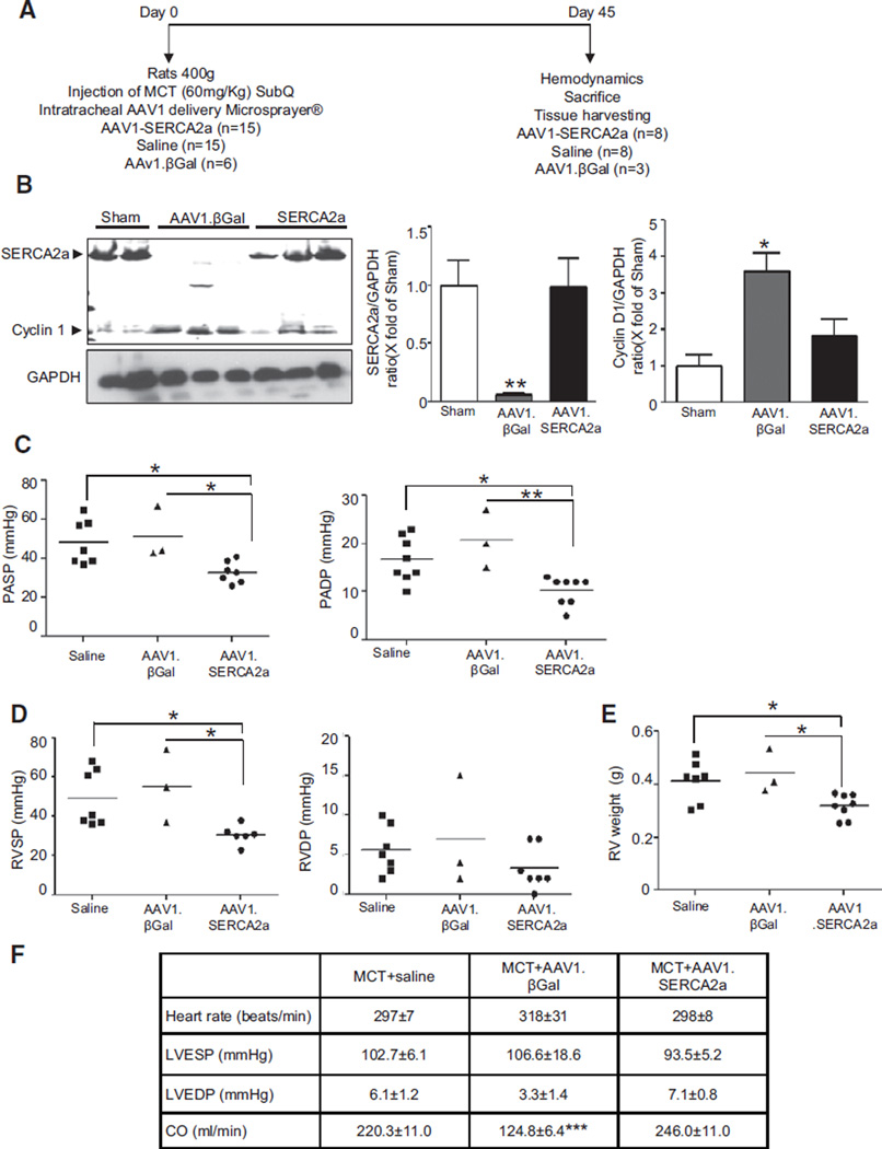
AAV1.SERCA2a prevents the development of PAH. A, Schematic of the protocol to assess the role of AAV1.SERCA2a gene therapy in the prevention of MCT-induced PAH. On day 0, rats were injected with MCT and coadministered aerosolized saline (n=15), AAV1.βGal (n=6), or AAV1.SERCA2a (n=15). B, SERCA2a and Cyclin D1 protein expression was examined by Western blotting in lung homogenates (n=3). Protein expression was normalized to GAPDH. Representative blots are shown. *P<0.04 vs saline, AAV1.SERCA2a, **P=0.008 vs saline, AAV1.SERCA2a. C, AAV1.SERCA2a limited the increase in pulmonary artery systolic (PASP) and diastolic (PADP) pressures observed in control group rats. Hemodynamics were measured invasively at day 45. *P<0.04 vs saline, AAV1.SERCA2a, **P=0.002 vs AAV1.SERCA2a. D, SERCA2a overexpression also limited the increase in RV systolic (RVSP) and diastolic (RVDP) pressures at day 45. *P<0.03 vs saline, AAV1. SERCA2a. E, RV hypertrophy was determined by the RV/LV+septum weight ratio (Fulton index). *P<0.02 vs saline, AAV1.SERCA2a. F, SERCA2a overexpression had no effect on LV systolic (LVSP) and diastolic (LVEDP) pressures or cardiac output (CO). ***P<0.001 vs MCT+saline, MCT+AAV1.SERCA2a. LV indicates left ventricle; MCT, monocrotaline, PAH, pulmonary arterial hypertension; and RV, right ventricle.

