
Figure 1.
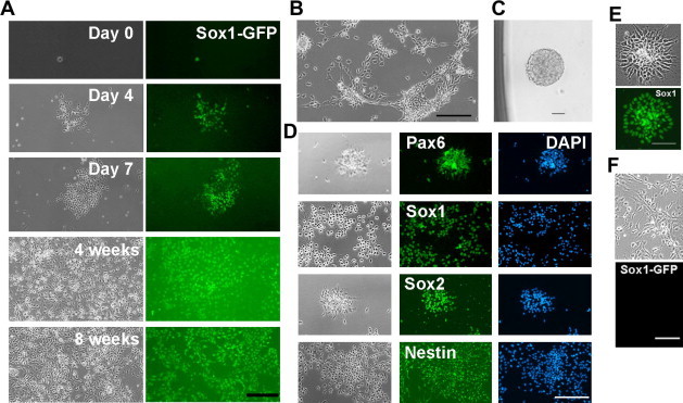
Expansion of Sox1-positive NS cells in the presence of LIF/CHIR/Y A. Clonal expansion of Sox1-GFP NS cells in the presence of LIF/CHIR/Y. Sox1-GFP positive NS cells derived from 46C ES cells were deposited into 0.1% gelatin-coated 96-well plates at 1 cell/well and cultured in N2B27 medium supplemented with LIF/CHIR/Y. Scale bar, 50 μm. B. Phase contrast image of NS cells derived from an E11.5 rat embryo and maintained in the presence of LIF/CHIR/Y at Passage 7. Scale bar, 50 μm. C. A neurosphere generated from a single primary rat NS cell in the presence of LIF/CHIR/Y. D. Immunostaining of primary rat NS cells maintained in the presence of LIF/CHIR/Y for 5 passages. Scale bar, 50 μm. E. Sox1 immunostaining of NS cells derived from H9 human ES cells and maintained in the presence of LIF/CHIR/Y. Scale bar, 50 μm. F. NS cells derived from 46C ES cells and maintained in the presence of FGF2/EGF for 2 passages. Scale bar, 50 μm.