
Figure 3.
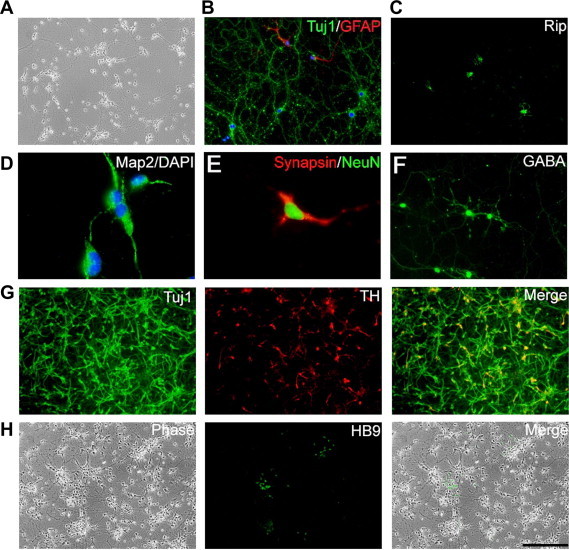
Neuronal and glial differentiation of rat iNS cells A. Phase contrast image of neurons spontaneously differentiated from rat iNS cells after the removal of LIF/CHIR/Y. B. Tuj1 and GFAP immunostaining of cells generated from rat iNS cells after exposure to EGF and FGF2 for 10 days followed by culturing in N2B27 medium plus 1% serum for another 7 days. C. Exposure to PDGF-AA and T3 (triiodothyronine) induced differentiation of rat iNS cells toward Rip-positive oligodendrocytes. D–H. Different subtypes of neurons derived from rat iNS cells. Scale bar, 50 μm.