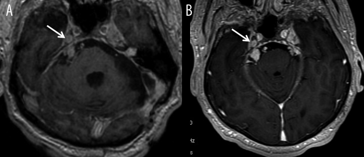
Figure 1.

Patient with type 2 neurofibromatosis, (A, B) – axial T1-weighted images after contrast administration. Image (A) was acquired few minutes after intravenous contrast injection while image (B) 10 minutes after injection of double dose of contrast agent. Image (B) shows significant improvement in detection of nerve V neuromas within the right cerebellopontine angle and Meckel’s cave (arrows).
