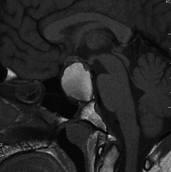
Figure 11.

A 21-year-old patient with a solid/cystic craniopharyngioma, located in the sellar-suprasellar region. Sagittal T1-weighted image shows high signal intensity of the cystic portion of the tumor as well as a significant enlargement of sella turcica and compression of the optic chiasm.
