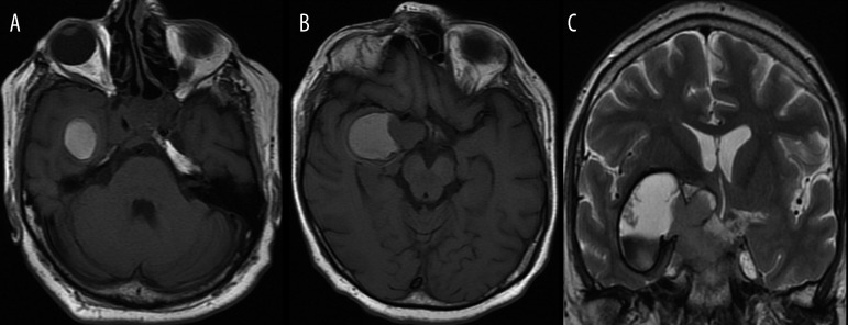
Figure 2.
Solid/cystic pituitary macroadenoma of prolactinoma type with hemorrhage during therapy with bromocriptine.
(A, B) – axial unenhanced T1-weighted images show high signal corresponding to methemoglobin.
(C) – coronal T2-weighted image allows for differentiation of methemoglobin types. The lower part of the tumor contains hypointense intracellular methemoglobin and the upper part of a lesion contains hyperintense extracellular methemoglobin.

