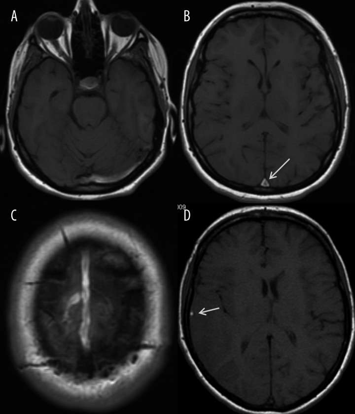
Figure 4.
Cerebral venous thrombosis, axial T1-weighted images. (A) – left sigmoid sinus thrombosis, (B) – superior sagittal sinus thrombosis in the inferior-posterior portion (arrow), (C) – superior sagittal sinus thrombosis at the convexity with a thrombosed draining cortical vein, (D) – thrombosis of the right vein of Labbe (arrow).

