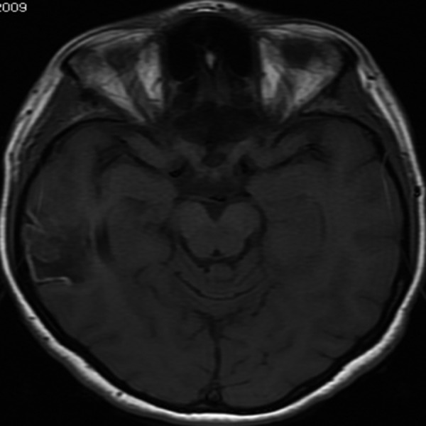
Figure 8.

Cortical laminar necrosis. Axial T1-weighted image, transverse section, demonstrates segmental necrosis of cerebral cortex visible as linear bands of high signal intensity in the right temporal cortex at the periphery of a chronic ischemic lesion.
