Figure 3.

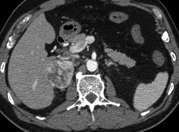
Axial CT scan after i.v. contrast enhancement – arterial phase (30 s), showing heterogeneous high contrast enhancement in the right adrenal mass that could be suspicious for pheochromocytoma.

Axial CT scan after i.v. contrast enhancement – arterial phase (30 s), showing heterogeneous high contrast enhancement in the right adrenal mass that could be suspicious for pheochromocytoma.