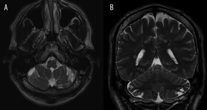
Figure 2.
(A) Patient 1: Axial TSE T2-weighted section. Presence of some cerebellar small cortico-subcortical longitudinal areas, isointense with CSF, orientated along the sulcis. (B) Patient 1: Coronal TSE T2-weighted section: Short, thin, atrophic folia of the superior semilunar lobules with dilatation of CSF spaces. Note the little periventricular hyperintensities of the supratentorial white matter.

