
Figure 4.
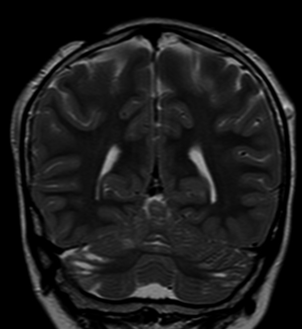
Patient 2: Coronal TSE T2-weighted section. Small CSF areas bilaterally located on the surface of the superior and inferior semilunar lobules. A left parietal subcutaneous vascular ectasia and a zone of aplasia cutis in the right parietal region are also evident.