
Figure 2.
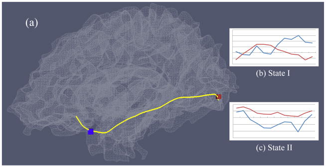
(a) Example of a SCGM voxel pair shown in red and blue boxes that are connected by DTI-derived fibers (in yellow). (b) The fMRI time series from the two voxels have low correlation within a specific time window (State I). (c) The fMRI time series from these two voxels are relatively higher correlated within another time window (State II).